Analytics: Royal Microscopical Society
2025 RMS Section Awards: winners announced!
Society recognises outstanding achievements in microscopy, imaging and flow cytometry
The Royal Microscopical Society (RMS) is pleased to announce the winners of its Science Section awards for 2025.
The awards, which are given out every two years, celebrate outstanding scientific achievements across all areas of microscopy and flow cytometry, with each RMS Science Section selecting a winner.
RMS President Peter O’Toole said: “It is a real privilege to announce our award-winners, who have all made some incredible contributions across their various scientific fields. Each winner has been chosen by one of our Scientific Committees – or ‘Sections’ – which represent all branches of microscopy, imaging and flow cytometry. As such, these awards are about receiving recognition from others working in the same field – and there’s no higher praise than that.
“The nominations were of exceptional quality and our committees certainly had a difficult job making their final decisions. My warmest congratulations go to all our winners.”
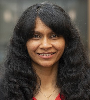
AFM and SPM Award:
Professor Sohini Kar-Narayan,
University of Cambridge, UK
Sohini has made outstanding contributions to the field of scanning probe microscopy (SPM) through her studies of nanoscale electromechanical properties of novel functional polymer and semiconducting nanostructures. Currently professor of Device Materials in the Department of Materials Science at the University of Cambridge, UK, she is internationally renowned for her pioneering research on functional materials for energy and biomedical applications. Her research group also introduced a novel time-resolved, open-circuit conductive atomic force microscopy (cAFM) technique as a new SPM methodology for direct electromechanical characterisation of semiconducting nanomaterials. Sohini is a world-renowned expert in the use of advanced SPM modes to understand structure-property and functionality relationships in novel polymer-based piezoelectric nanostructures. She has pioneered the use of piezoresponse force microscopy (PFM), Kelvin probe force microscopy (KPFM) and quantitative nanomechanical mapping (QNM) to reveal the electromechanical properties and lamellar structure of a range of different piezoelectric polymers at the nanoscale.
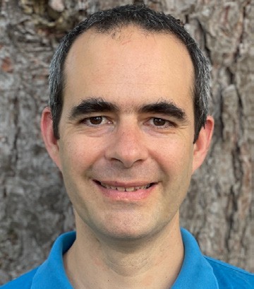
Award for Data Analysis in Imaging (DAIM):
Dr Francisco de la Peña,
University of Lille, France
Francisco is a researcher in the fields of analytical electron microscopy, materials science and scientific data analysis. As the primary creator of HyperSpy, a python package for the analysis of multidimensional data, with specific functionality for analysing data collected in electron microscopes, he has made an unparalleled contribution to the analysis of electron microscopy data in physical sciences. Francisco is also a regular speaker at a number of conferences and workshops promoting open-source software development for electron microscopy. Arguably no other individual has impacted the analysis of electron microscopy data in the physical sciences as much as Francisco over the last decade and a half.
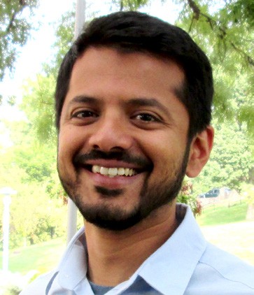
Alan Agar Award for Electron Microscopy: Dr Kedar Narayan, Frederick National Laboratory, National Cancer Institute, NIH, Maryland, US
Kedar has been among the leading figures in the development of Volume Electron Microscopy – a high-end EM technique – for several years. He is a senior scientist and group leader at the Center for Molecular Microscopy (CMM) at Frederick National Laboratory and National Cancer Institute, NIH, Maryland, US. He earned a PhD in immunology, with an emphasis on biophysics and imaging, at the Johns Hopkins School of Medicine in 2008. He also has a background in chemistry, pathology and software engineering. Since 2015 at the CMM – a highly collaborative laboratory with a strong technology development portfolio – his group has developed and applied FIB-SEM and other volume EM technologies to questions in cell biology.
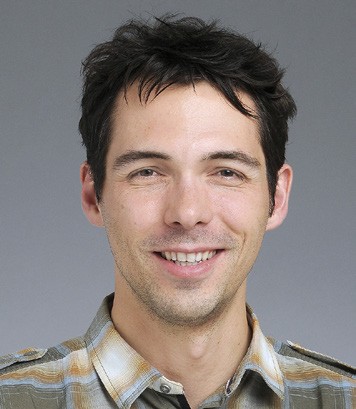
Award for Flow Cytometry:
Dr Florian Mair,
Eidgenössische Technische Hochschule (ETH), Zurich, Switzerland
Florian has made a number of outstanding contributions in the field of cytometry during a relatively short period of time. His journey from PhD and postdoc, to running a Core facility, has been a remarkable one, in which cytometry has been pivotal. Dr Mair completed his PhD at the University of Zurich, Switzerland, in 2013 and did postdoctoral research there while also working within the University’s Flow Cytometry Facility. In 2017, he moved to the Fred Hutchinson Cancer Research Institute in Seattle, Washington, US, in the Lab of Martin Prlic, which looks at T-cell responses in inflammation. While in the Prlic Lab, he developed the first 28 colour fluorescence panel to look at human dendritic cells. At the time, this was the maximum number of analytes that could be measured. In May 2022, he moved back to Switzerland to become scientific director of the flow cytometry facility at the ETH in Zurich.

Award for Innovation in Applied Microscopy for Engineering, Physical and Material Sciences:
Professor Emilie Ringe,
University of Cambridge, UK
Emilie is a stand-out materials researcher, utilising both optical and electron microscopy for developing new, optically active nanomaterials and establishing new materials for light-assisted catalysis. Having carried out seminal work in plasmonic nanoparticles in her PhD at Northwestern University, Illinois, US, Emilie has pioneered Wulff-construction type principles and user-friendly codes for twinned, alloyed and kinetically controlled crystal shape prediction. She has pioneered the synthesis of well-defined, air-stable plasmonic magnesium nanocrystals. Her work on plasmonics has made remarkable use of monochromated electron energy loss spectroscopy in the scanning transmission electron microscope (STEM-EELS) for extracting spectra and maps of the plasmonic near-field response characteristics. Emilie has been an active developer of novel optical microscopy techniques, including hyperspectral and compressive methods for optical imaging and spectroscopy.

Award for Life Sciences: Dr Vaishnavi Ananthanarayanan, University of New South Wales, Australia
Vaishnavi has made a number of remarkable contributions to the field of Cell Biology, harnessing wide-ranging microscopy techniques and modalities. She currently holds a nine-year EMBL Australia Group Leader fellowship and leads a large research group at UNSW. As a leader in the research fields of motor proteins and their regulation; the roles of the cytoskeleton in organelle remodelling and organisation; and cellular decision-making, Vaishnavi has been one of the true pioneers in quantitative single molecule microscopy to understand the biophysical principles of these processes. Her research has consistently featured novel imaging methods such as live-cell single-molecule imaging, and technologies like optogenetics, microfluidics and micropatterning. She is also a true champion of equity and inclusion, having organised a number of research-specific as well as inclusion-focused events, and is co-founder of BiasWatchIndia, an initiative to document women’s representation in Indian scientific conferences.
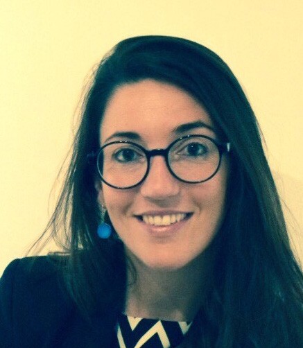
Award for Light Microscopy:
Dr Ilaria Testa,
KTH Royal Institute of Technology, Stockholm, Sweden
Ilaria is an internationally recognised multidisciplinary researcher who has made outstanding contributions to the field of super-resolution optical imaging. She started her own group – SciLifeLab – in 2015, following the award of a competitive ERC Starter fellowship at the KTH Royal Institute of Technology in Stockholm, Sweden, where she now holds an Associate Professorship. She has led her team in the creation of new and exciting fluorescence nanoscopy technologies aimed at improving the speed and resolution of the light microscope, while remaining sufficiently gentle for live cell imaging. Ilaria’s application of innovative super-resolution microscopy methods in cell biology has been particularly impressive.